Aside from being a thing of beauty, the feline eye is an important, delicate organ. Cats are predators, and their eyesight has evolved to assist them in hunting. As nocturnal creatures, cats are more sensitive to light. While they can’t see in total darkness, cats require only one-sixth the amount of light as that of a person to see. Their pupils can dilate three times larger than a human’s, and the cornea is bigger, allowing for more light to enter.
Occasionally, cats develop a problem with one or both eyes, especially the corneas. Anyone who has ever experienced having an eyelash trapped under a contact lens or a grain of sand blown into their eye quickly discovers that the cornea is teeming with pain receptors. A corneal ulcer — a scratch or scrape involving the cornea — is an uncomfortable, potentially vision-threatening disorder in cats.
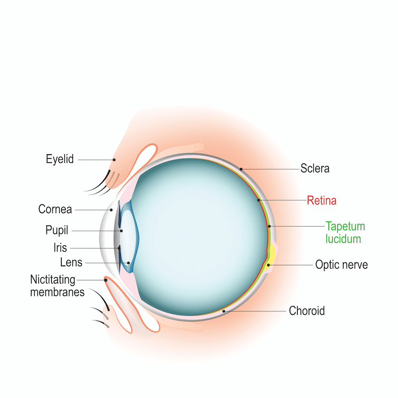
What is the cornea?
The cornea is the clear membrane that covers the surface of the eyeball, essentially acting as a windshield. It is composed of several layers. The outer surface is called the epithelium. Just beneath the epithelium is the stroma. The innermost layer is called Descemet’s (pronounced “dessa-mets”) membrane.
Cats have developed well-refined mechanisms to prevent damage to their corneas. They have vibrissae (those big “whiskers” above their eyelids), which are good at detecting objects that approach their eyes, allowing them to take evasive action. Cats also have a well-developed blink response. In addition, they have a muscle attached to the back of their eyeball (the retractor bulbi muscle) that can pull the eyeball back into the socket. This allows a membrane (the nictitating membrane or “third eyelid”) to elevate, protecting the cornea.
Despite these sophisticated mechanisms, cats occasionally suffer trauma to the cornea, and an erosion or abrasion occurs on the corneal surface. As noted above, we call this open wound within the cornea a corneal ulcer. Most ulcers involve the surface epithelium. Some ulcers go a little deeper, into the stroma. If it goes further into the stroma all the way down to Descemet’s membrane, the ulcer is called a descemetocele (pronounced “dessa- meta-seal”), a perilous situation with little leeway. If the ulcer goes deeper, through Descemet’s membrane, the eyeball will rupture and vision will be lost.
The most frequent cause of corneal ulcers in cats is infection with the feline herpesvirus. Trauma is another common cause, such as a scratch from another cat, rubbing the eye on the carpet or an unexpected interaction with a plant or tree branch. Foreign bodies and chemicals can also abrade the cornea, but these are less likely scenarios. Eyelid disorders are another possible cause of corneal trauma. Entropion is a condition in which the eyelid rolls inward, causing hair near the margin of the eyelid to contact the cornea. Over time, this can lead to an ulcer. I have surgically repaired many cases of entropion. Any breed of cat may acquire a corneal ulcer, but breeds with bulging eyes, such as Persians, are at increased risk.
Diagnosis
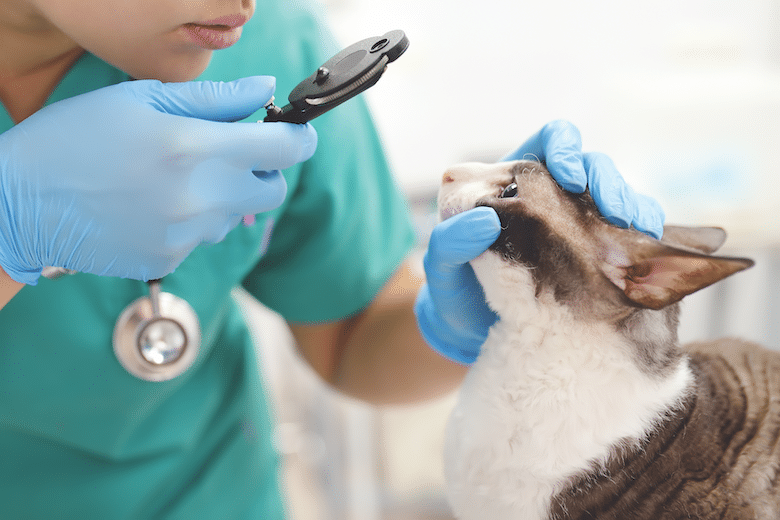
Because corneal ulcers are painful, most affected cats will show signs of discomfort, such as tearing, rubbing the eye and keeping the eye partly or completely closed. To prove that an ulcer is the cause of the discomfort, a fluorescein stain is usually performed. To perform this test, a drop of a fluorescent orange-colored liquid is applied to the cornea. If the cornea is intact, the dye washes smoothly over the corneal surface. If an erosion or ulcer is present, however, the dye will adhere to the ulcerated area and can be easily detected using a black light.

Treatment
Regardless of the cause, corneal ulcers must be treated promptly. The feline cornea is only 0.5 millimeters thick. Delaying therapy can lead to rupture of the eyeball and loss of vision.
Treatment varies, depending on the depth and severity of the ulcer. Antibiotic drops or ointment is applied to the cornea several times a day to prevent an infection from occurring.
How to administer eye medication
Treatment for corneal ulcers involves administering drops or ointments. Drops are often easier to administer. Ointments have the advantage of providing lubrication and allowing for increased contact time for the medication and are especially useful given at bedtime. Ointment application involves using the thumb or forefinger to gently roll the lower eyelid downward. Ointment is then squeezed into the exposed space (called the “conjunctival sac”), and the eye is opened and closed by hand several times to evenly distribute the ointment over the eye. Approaching the eye from the outside corner can prevent the cat from seeing the tip of the tube, making administration a bit easier. Eye drops are instilled with the cat’s nose tilted slightly upward. To prevent contamination, the tip of the dropper bottle or ointment tube should not be touched by fingers or any other surface, and should not come into direct contact with the eye.
Irritation of the cornea often leads to spasm of a muscle inside the eye called the ciliary muscle. When this muscle spasms, it causes pain for the cat. Atropine drops or ointment applied to the affected eye causes paralysis of the ciliary muscle, reducing pain and discomfort. Atropine will cause the pupil to dilate widely, making the affected eye very sensitive to light and causing squinting, especially in bright light. Cats who rub at their eye a lot may need to wear an Elizabethan collar to prevent further trauma. If the herpesvirus is the suspected cause, anti-viral medicine is warranted. Ulcers caused by the herpesvirus typically take longer to heal than superficial ulcers caused by trauma.
Superficial ulcers typically heal in three to five days. After a few days of ulcer treatment, the fluorescein stain test is performed again. If the cornea does not take up any stain, it is considered to be healed.
Deep ulcers that are at risk for perforating require more aggressive therapy, such as applying a special soft contact lens to the affected cornea or some type of surgical technique designed to cover the ulcer. A common surgical procedure is a conjunctival graft. In this procedure, a small piece of tissue adjacent to the cornea is sutured over the ulcer. This allows blood vessels to deliver nutrients, antibodies and infection-fighting cells to the damaged cornea, as well as providing mechanical support, in a similar fashion to how a skin graft promotes healing of a severe burn. Complicated cases like this require referral to a board-certified veterinary ophthalmologist.
A corneal ulcer is a potentially vision-threatening condition. Prompt diagnosis and aggressive treatment is required to preserve vision and keep cats comfortable.
Top photograph: SbytovaMN | Getty Images
Read Next: Cat Pupils: Here’s What Your Cat’s Eyes Are Telling You

